3D tomography and diagnostics
Cone-beam 3D CT, panoramic X-ray (orthopantomogram) and radiovisiography.
About the treatment
Three-dimensional computed tomography (CT) is the gold standard of modern dental radiodiagnostics.
A latest-generation cone-beam scanner produces a 3D image of both jaws at the highest resolution and with a minimal radiation dose.
CT lets the dentist assess the volume and density of bone for an implant in 3D, and check the position of the root canals, the sinuses and the trigeminal nerve.
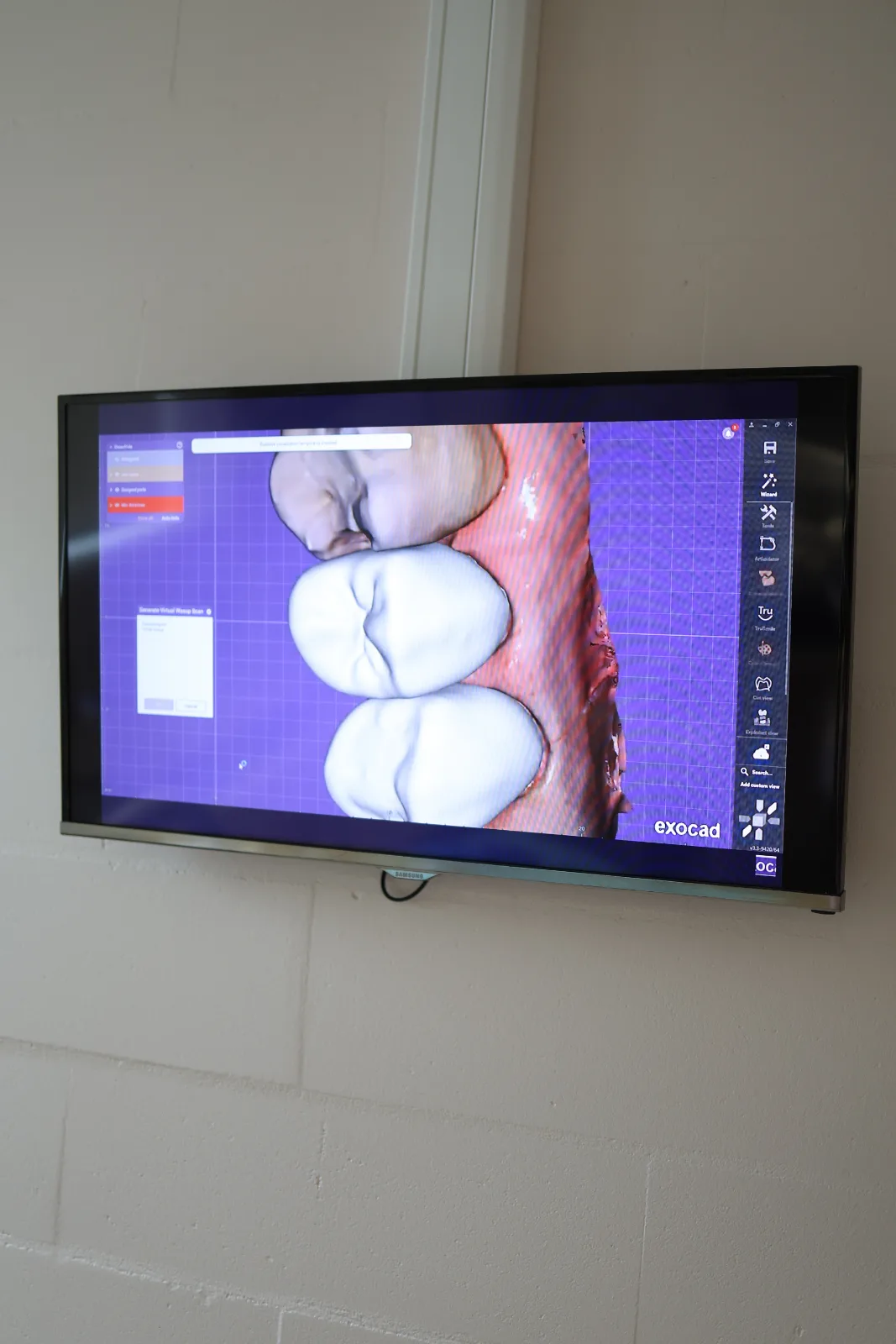
Besides the scanner, the room has an intraoral scanner. It takes a digital impression of the jaw in a few minutes — no tray, no impression material, and therefore no gag reflex. The finished model goes straight to the laboratory or into the planning software.
Zebris axiography records how the lower jaw actually moves: while chewing, opening and shifting sideways. The bite is adjusted from that recording. It is needed when extensive prosthetic work is planned or when the joint clicks and hurts.
What the visit includes
- Minimal radiation dose
- 3D scan of both jaws, can be saved to digital media
- 2D panoramic X-ray
- Result ready in 2 minutes
- Intraoral scanner instead of impressions
- Zebris axiography: digital bite registration and computer analysis
When this procedure is needed
- An implant is planned: the volume of bone and the position of the nerve need to be known.
- A wisdom tooth lies horizontally or close to the mandibular canal.
- There is pain but an ordinary X-ray shows nothing.
- Orthodontic treatment or complex prosthetic work is planned.
- An impression is needed for a crown, a splint or aligners — we take it with a scanner, with no tray in your mouth.
- The jaw joint clicks or hurts: axiography shows how it actually moves.
How the visit goes
- 1PreparationEarrings, glasses and anything metal are removed from the field. The procedure needs neither anaesthesia nor preparation the day before. Women are asked to tell us about a possible pregnancy before the procedure. Computed tomography is contraindicated for children under 10.
- 2The scanYou stand still for 15–20 seconds while the machine makes a turn around your head. The radiation dose is comparable with a few hours of air travel.
- 3Report and handoverThe dentist goes through the scan on screen with you. The file is given to you on a disc or sent as a link — you can show it to any other dentist.
Prices for this service
- Single-tooth X-ray (radiovisiography)20 €
- Panoramic X-ray of both jaws (OPG)28 €
- 3D computed tomography, one quadrant50 €
- 3D computed tomography, one jaw40 €
- 3D computed tomography, both jaws76 €
- Intraoral scan (digital impression)35 €
- Zebris axiography: digital bite registration and computer analysis120 €
Treatment covered by Tervisekassa
For patients with a confirmed severe disability or special mental health needs, and for children up to 19 (where indicated), Tervisekassa covers this treatment — you pay nothing at the clinic. Ask reception what applies in your case.
After the procedure
- The scans stay at the clinic on a strictly protected server. On request we can upload them to Pildipank, though there they are unfortunately stored in a reduced form. For a small fee they can be saved to digital media. CT scans are too large to send by e-mail; other images are sent only in encrypted form upon a written request.
- The data holds for about six months to a year. If teeth have been removed or treated meanwhile, a fresh scan is taken before surgery.
- There are no restrictions afterwards: you can eat straight away, drive and go to another appointment.
- The digital impression stays at the clinic. For repeat prosthetic work a new scan may not be needed.
Frequently asked questions
Is computed tomography harmful?
The dose is comparable with a few hours of air travel. A scan is taken when there is a reason, not "just in case" — that is the main protection.
Can I bring a scan from another clinic?
Yes, if it is no more than six months to a year old and comes on an external digital carrier or as a link. The dentist will look and say whether it is enough or a fresh one is needed. Because of differing standards a scan may also fail to open in our system — then a new one will be needed after all.
How does a digital scan differ from an impression?
The scanner captures the shape of the teeth with a camera — no tray, no impression material. It is more accurate and there is no gag reflex.